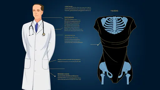
The human rib cage, a complex structure of bones and cartilage, serves as a protective fortress for vital organs. Among its many fascinating aspects, the phenomenon of costal flaring—often colloquially referred to as "flared ribs"—has garnered attention in both medical and fitness communities. This condition, where the lower ribs protrude outward abnormally, can sometimes be detected through X-ray imaging, revealing subtle or pronounced deformities in the thoracic architecture. While some cases are congenital, others develop due to postural habits, muscular imbalances, or underlying skeletal disorders. The visual evidence provided by X-rays offers clinicians a critical tool for diagnosis and treatment planning.
Radiographic imaging plays a pivotal role in assessing rib cage abnormalities. When a patient presents with symptoms such as chronic discomfort or restricted breathing, an X-ray can reveal whether the rib distortion is structural or positional. For instance, flared ribs may appear as an outward splay of the lower ribs, often asymmetrical, with potential secondary effects on spinal alignment. In contrast, "sunken rib" deformities, another variant, might show an inward collapse of the costal margin. These images help differentiate between benign anatomical variations and pathological conditions like scoliosis or thoracic insufficiency syndrome.
The causes of rib cage deformities are multifaceted. Prolonged poor posture, particularly in sedentary individuals, can contribute to muscular weakness around the thorax, allowing the ribs to shift over time. Athletes, especially those in sports requiring repetitive rotational movements (e.g., golfers or baseball pitchers), may develop unilateral rib flaring due to uneven muscle development. Additionally, connective tissue disorders such as Ehlers-Danlos syndrome can lead to hypermobility of the rib joints, exacerbating distortions. X-rays not only confirm these diagnoses but also track progression, informing decisions about bracing, physical therapy, or surgical intervention.
Beyond clinical implications, the aesthetic and psychological impact of flared ribs shouldn’t be underestimated. Many individuals seek medical evaluation after noticing a protruding rib contour, particularly in fitness settings where torso definition is emphasized. X-rays provide objective data to distinguish between actual skeletal distortion and superficial concerns exacerbated by low body fat. This distinction is crucial, as "corrective" exercises for purely cosmetic concerns may be unnecessary or even harmful if misinterpreted without imaging.
Emerging technologies are refining how we analyze rib deformities. While traditional X-rays offer static snapshots, dynamic imaging techniques like fluoroscopy capture real-time rib movement during respiration. This is particularly valuable for assessing functional impairments linked to flared ribs, such as reduced diaphragm efficiency. Furthermore, 3D reconstructions from CT scans provide unparalleled detail for surgical planning in severe cases. Yet, despite these advances, the humble X-ray remains a frontline diagnostic tool due to its accessibility and cost-effectiveness.
Management strategies vary widely based on radiographic findings. For mild, asymptomatic flared ribs, observation and postural retraining may suffice. More pronounced deformities affecting respiratory function might require targeted physical therapy to strengthen the intercostal and core muscles. In rare cases where organ compression or severe deformity exists, surgical options like rib resection or stabilization become considerations. Here, pre- and post-operative X-rays are indispensable for evaluating anatomical changes and surgical outcomes.
The intersection of radiology and biomechanics continues to deepen our understanding of rib cage dynamics. Research utilizing X-ray motion analysis has revealed how even subtle rib distortions alter load distribution across the thorax, potentially contributing to chronic pain patterns. This knowledge informs more nuanced rehabilitation approaches, moving beyond generic "core strengthening" to address specific muscular deficits identified through imaging. As awareness grows, so does recognition that rib cage morphology exists on a spectrum, with "normal" encompassing considerable variation.
Patient education is a critical component of managing rib-related concerns. Clear communication of X-ray findings helps individuals understand whether their flared ribs represent a structural anomaly or a functional adaptation. This distinction guides realistic expectations for interventions—whether aiming to improve posture, alleviate discomfort, or address athletic performance limitations. Radiologists increasingly collaborate with physiotherapists to create tailored management plans, bridging the gap between imaging diagnostics and practical therapeutics.
Looking ahead, advancements in low-dose radiographic techniques promise safer longitudinal monitoring of rib cage development, particularly in pediatric populations where early detection of progressive deformities is crucial. Simultaneously, machine learning applications are being trained to detect subtle rib abnormalities that might escape the human eye, potentially flagging early signs of systemic connective tissue disorders. Yet, amidst these technological strides, the clinician’s interpretive expertise remains irreplaceable—correlating imaging findings with the patient’s unique history and symptoms.
Ultimately, the study of rib cage deformities via X-ray imaging exemplifies the marriage of structural anatomy and functional medicine. From diagnosing rare syndromes to optimizing athletic performance, these radiographic insights illuminate a bony framework that is anything but static. As research unravels the rib cage’s nuanced role in overall biomechanics, the humble rib X-ray stands as a testament to how foundational imaging technologies continue to evolve in their diagnostic and therapeutic relevance.

By /Aug 6, 2025

By /Aug 6, 2025

By /Aug 6, 2025

By /Aug 6, 2025

By /Aug 6, 2025

By /Aug 6, 2025

By /Aug 6, 2025

By /Aug 6, 2025

By /Aug 6, 2025

By /Aug 6, 2025

By /Aug 6, 2025

By /Aug 6, 2025

By /Aug 6, 2025

By /Aug 6, 2025

By /Aug 6, 2025

By /Aug 6, 2025

By /Aug 6, 2025
By /Aug 6, 2025

By /Aug 6, 2025

By /Aug 6, 2025